
Inspection
- Assess posture, gait pattern, how the patient sits/stands up from a chair, whether back movement triggers pain, and whether the patient experiences muscle weakness in the lower extremities.
- Consider to determine if there are normal axes and curves in the back, scoliosis, pelvic drop, and normal paraspinal musculature.
Thoracic
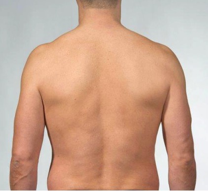
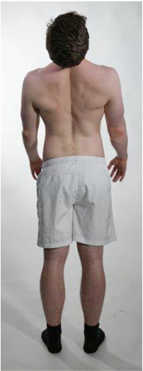
Inspection from behind. Look for axes and curves in the back. Note signs of atrophy or hypertrophy in the back muscles. Note if there are signs of scoliosis. Note if there is normal thoracic kyphosis.
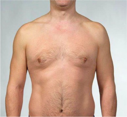
Inspection from the front. Look for symmetry in the thorax. Note if there are signs of atrophy or hypertrophy in the chest muscles.
The spinous process of C7 can be found on a straight line from the acromion towards the spine, Th3 on a straight line from the scapular spine towards the spine, and Th7 on a straight line from the scapular angle towards the spine.
Lumbar
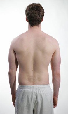
Inspection from behind. Look for axes and curves in the back. Note signs of atrophy or hypertrophy in the back muscles. Note if there are signs of scoliosis. Note if there is a normal lumbar lordosis.
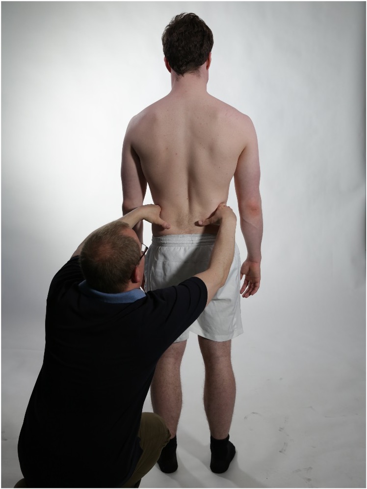
Check pelvic position and note if there is a pelvic tilt.
Level Th12 is found in the middle of the line between the scapular angle and the iliac crest. The spinous process L4/L5 is found on a straight line from the iliac crest, and the spinous process L5/S1 is found on a straight line between the dimples.
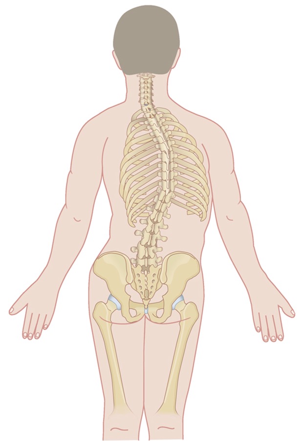
Thoracic scoliosis
Lumbar scoliosis
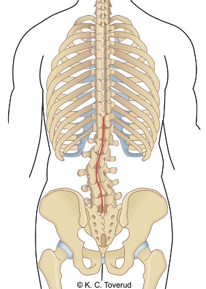
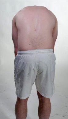
Assess whether there are signs of structural scoliosis. Be particularly attentive to this when examining adolescents with back pain. The aim of the examination is to diagnose the condition, and based on clinical findings, to assess whether further investigation is necessary. Structural scoliosis is characterized by a gibbus (rib hump) when bending forward, often accompanied by pelvic tilt and asymmetry in the shoulders and iliac crest. A first step in the investigation is to refer for scoliosis X-rays to clarify the size of Cobb's angle. The value of Cobb's angle determines how the patients should be followed up.
Cobb's angle
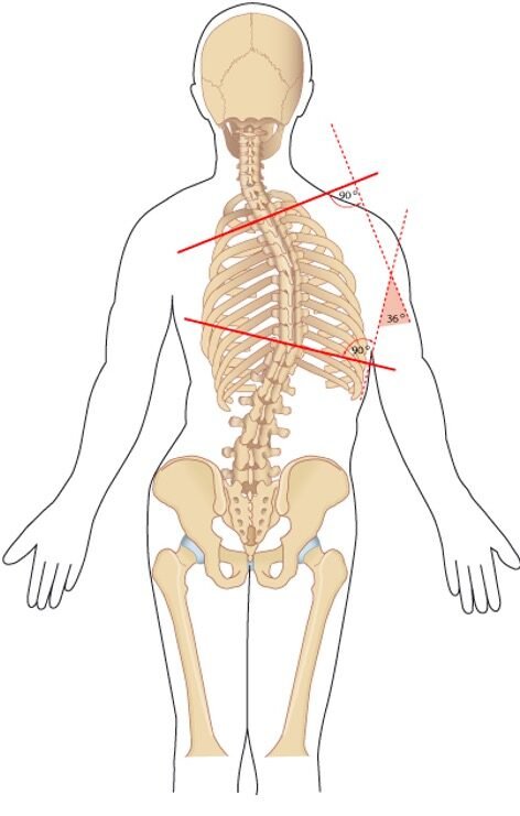
The Cobb angle is measured by drawing two parallel lines from the two most tilted vertebrae in the scoliosis curve and erecting intersecting lines perpendicular to these two parallel lines. The angle formed between the two parallel lines and the perpendicular lines is called the Cobb angle.
Cobb angle and treatment of scoliosis
- Cobb angle < 25 degrees: Usually no treatment, observation with check-ups every 6 months during remaining growth
- Cobb angle > 25 degrees and remaining growth: Usually brace treatment
- Cobb angle > 45-50 degrees: Usually surgery
- When fully grown, a scoliosis of < 50 degrees usually does not worsen
Increased thoracic kyphosis, consider Scheuermann's disease
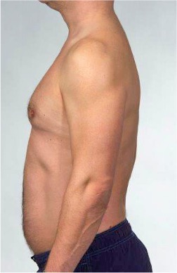
With increased thoracic kyphosis, one must consider whether Scheuermann's disease is present.
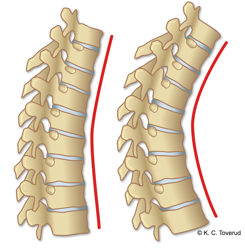
The condition is caused by a juvenile osteochondrosis. At least three wedge-shaped vertebrae (≥ 5°) in the thoracic spine increase kyphosis. It most often occurs in boys between 13 and 16 years old, usually towards the end of the growth period. Most have little pain or discomfort.
Active movements
- Assess the patient's ability and willingness to move the back. Record the range of motion, and ask if the movements trigger back pain or radiating pain to the lower extremities.
- Consider whether extension (25–45°), flexion (thoracic 20–45°, lumbar 80–90°), sideways bending (20–40°), and rotation (45°) cause back pain or radiating pain to the lower extremities. Observe whether the mobility of the back occurs smoothly, and note the distance between the fingers and the floor. The distance is measured in cm from fingertip to floor. There is great individual variation. Fingertip–floor distance > 10 cm may indicate limited mobility in the spine.
Extension
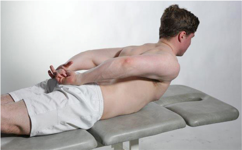
Central lumbar pain can be caused by facet joint osteoarthritis, pain from ligaments, or disc disorders, radicular radiation is often seen with stenosis in the neuroforamina which can cause compression of the exiting spinal nerve.
Thoracic flexion
Lumbar flexion
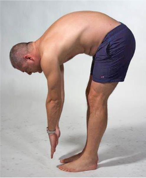
- Look for gibbus (rotational deformity), this is a typical finding in structural scoliosis. Viewed from the side, it is possible to assess whether there is smooth segmental mobility in the spine and, if relevant, how much of the flexion movement occurs at the hip joints.
Lateral flexion

Note how far down toward the knee joint the fingertips reach, and compare sides. Note any pain radiation in the lower extremities.
Rotation
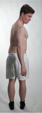
In a standing position, the rotational movements become a combined movement in the hip, lumbar spine, and thoracic spine. Note range of motion, any side differences, and whether the movement triggers pain.
Strength squat
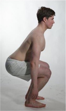
Screening test for strength knee flexion. Joint in an orienting neurological examination for radiculopathy in spinal nerve L2-L3.
Strength heel raise
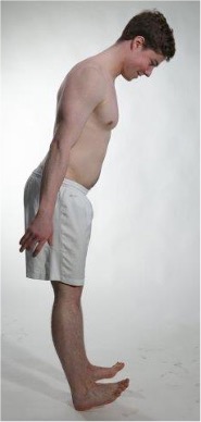
Screening test for strength heel walk. Part of an orienting neurological examination for radiculopathy in spinal nerve L4–L5
Toe-walking strength

Strength "TipToe"
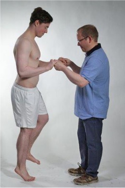
Screening test for strength toe walking and strength during single-leg heel raise. Part of an orienting neurological examination for radiculopathy in spinal nerve L5–S1
Trendelenburg test
Tested in single-leg stance
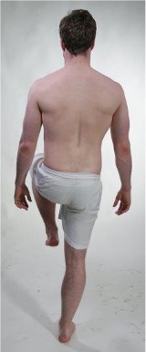
Negative Trendelenburg's test. The examiner asks the patient to stand on one leg. Normally, the pelvis on the lifted leg is kept in a straight line or raised slightly.
Positive Trendelenburg sign
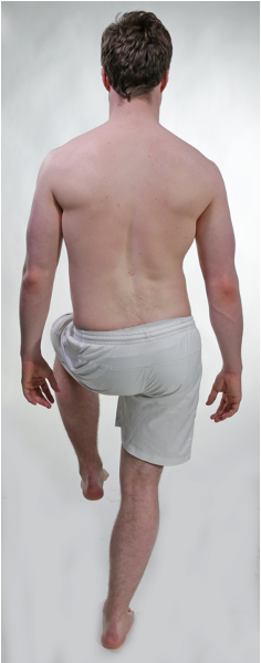
Positive Trendelenburg test.
With a positive test, the pelvis on the leg being lifted will drop.
Trendelenburg sign
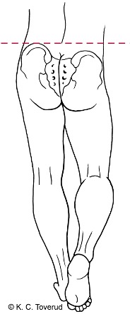
- A positive test is a sign of weakness in the gluteus medius muscle on the stance leg.
Thoracic excursion expiration
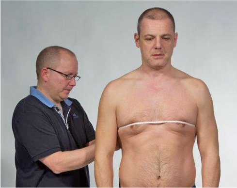
Expiration. Find the 4th intercostal space. Have the patient exhale, and measure around the horizontal plane.
Thoracic excursion inspiration, + 3 cm
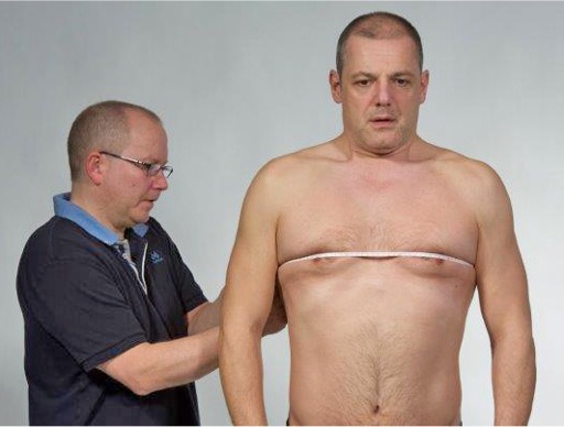
Inspiration. Have the patient inhale as much as he or she can, and measure again. The best of two attempts counts.
The test is used as part of the diagnostics when inflammatory spinal disease is suspected. Differences in circumference between inhalation and exhalation < 3 cm are pathological.
Passive movements
- Assess Passive rotation in the thoracic spine with the patient seated on a bench. Passive extension of the back is easiest performed with the patient lying prone. The patient is instructed to place their hands under their body and lift the upper body.
- Consider on the range of motion during rotation is symmetrical, whether there is pain at the end range of motion, and whether the pain radiates to the front of the chest or is localized in the back. Rotation with neck flexion can increase pain if there is a thoracic disc disorder. The pain then typically has a radiating character from the back toward the rib cage, often located on the side to which the back is rotated.
Starts by examining passive rotation in the thoracic spine with the patient sitting on a bench, assessing whether the range of motion is symmetrical, whether there is pain at the end range of motion, and whether the pain radiates to the front of the chest. Flexion in the neck can increase the perception of pain if there is a thoracic disc disorder.
Rotation 45°
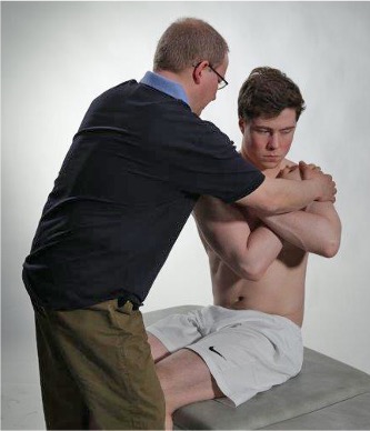
Pain at the end range of motion radiating from the back to the chest can be a clinical sign of thoracic disc disorder or facet joint osteoarthritis.
Rotation with neck flexion
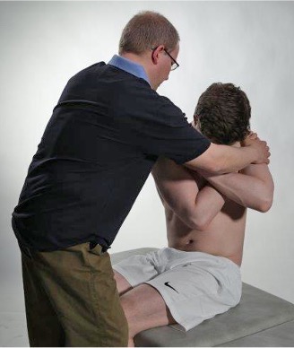
Rotation with neck flexion can increase pain if there is a thoracic disc disorder.
Extension 25-45°
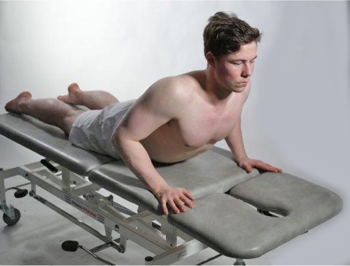
If passive extension in the back central back pain occurs, there may be facet joint osteoarthritis, disc disorder or ligamentary pain. If there is a narrow neuroformina, the nerve root can be pinched during this movement and cause radiating pain to the lower extremities. Make a note of the range of motion and whether the movement triggers pain.
Springing test thoracic
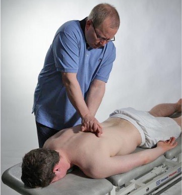
Spring test thoracically and lumbar is performed to check if there is local pain due to degenerative disorder in the facet joints or if pain comes from ligamentary structures.
Iliosacral joint compression
Iliosacra joint compression test and passive flexion in the hip joint are carried out as a screening and in the event of a negative test reduce the likelihood of low back pain coming from the pelvic joint or hip joint.

The examination is carried out with the patient in the supine position. The examiner has crossed hands on the spina iliac anterior superior. The test is used to clarify whether low back pain or pelvic pain can come from the iliosacral joint. If the iliosacral joint test reproduces the patient's pain, a full examination of the iliosacral joint proceeds.
Iliosacral joint compression
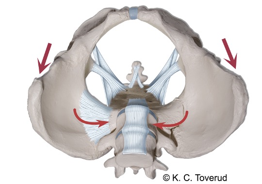
- The test involves compression of the sacroiliac joint posteriorly. The test has the potential to reproduce the patient's pain if it originates from an active sacroiliac joint arthritis. The examiner records whether the test triggers unilateral or bilateral pain over the sacroiliac joint.
Hip flexion
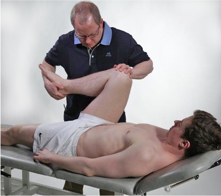
Flexion in the hip joint is used as a screening test. The test is used to determine if low back pain or pelvic pain may come from the hip joint. If passive flexion in the hip joint reproduces the patient's pain, a full examination of the hip joint is carried out.
Isometric movements
- Assess strength and any pain from composite muscle groups that provide movement in the body. It is important to have knowledge of the function of the large muscle groups in the body and back.
- Consider strength performance in the range of motion and pay particular attention to the fact that it is muscle groups being tested, not individual muscles.
Palpation
- Assess whether there is local pain, referred pain, or muscle tension.
- Consider where the pain comes from with palpation of relevant painful muscle groups to find a more accurate location, does the pain come from the interscapular muscle groups, the lower back, or from the trochanter area. Use palpation to confirm findings from active/passive/isometric testing. Pay particular attention to spondylolisthesis, iliolumbar pain, gluteus medius tendinopathy, and myofascial trigger points that cause referred pain.
Intercostal musculature
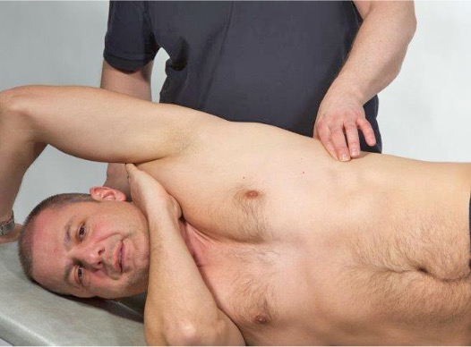
Very many adults are unable to breathe using the diaphragm, and raise the chest and shoulders when taking a breath in. This strain is something we are really not made to endure, and very many people therefore also experience pain in these muscles that lift the chest.
Upon examination, one usually finds a patient who complains of stabbing pain, there is localized muscle tension, and there is pronounced tenderness in the muscles when pressing on the chest in the painful area.
Intercostal musculature
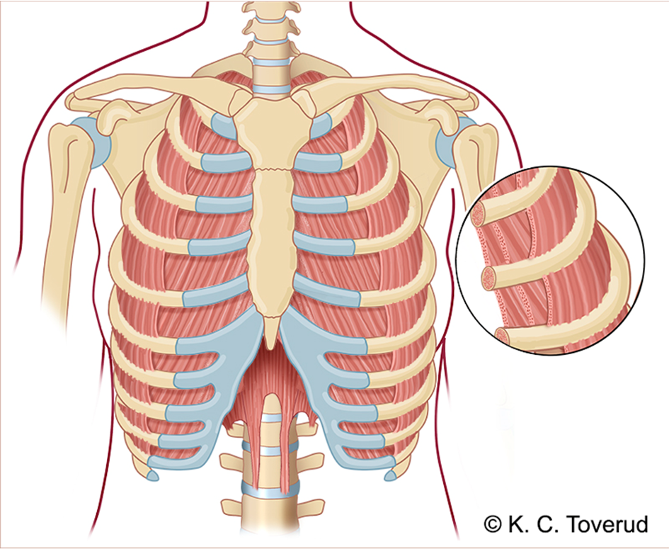
Mm. intercostales externi, N. thoracicus. Increases the volume of the thoracic cavity by pulling the ribs upwards – inhalation.
Mm. intercostales interni, N. thoracicus. Reduces the volume of the thoracic cavity by pulling the ribs downward – exhalation.
M. rhomboideus minor and major
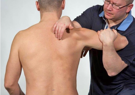
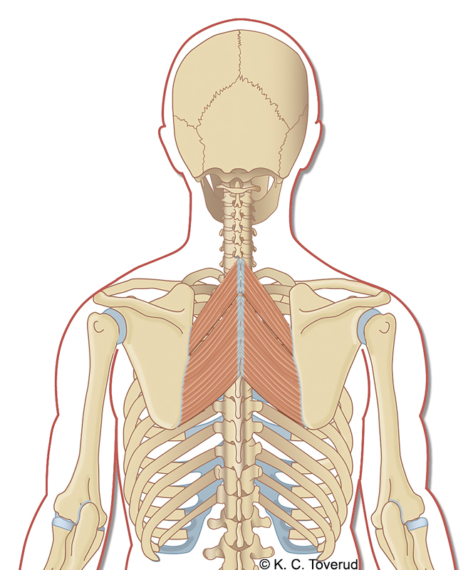
Tensions in the muscles cause interscapular pain, often associated with movement of the shoulder blades or in connection with breathing. On examination, tender muscle knots and crepitation are often detected when the scapula moves.
Spinous process
Step formation?
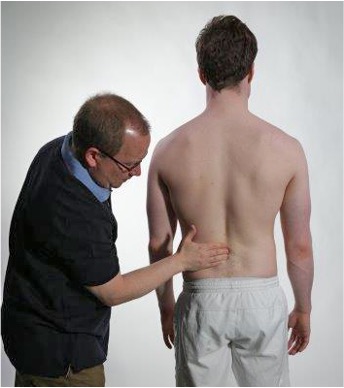
Palpate along the spinous processes while the patient is standing. Feel for a height difference (step-off) where you notice that a spinous process appears to be shifted forward. Such a finding may be consistent with spondylolysis, and as a consequence of this, spondylolisthesis.
Pain?
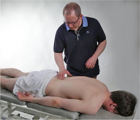
With the patient in the prone position, the examiner palpates along the interspinous ligament. Note if palpation triggers pain, and at which disc level the pain is located.
Spondylolisthesis
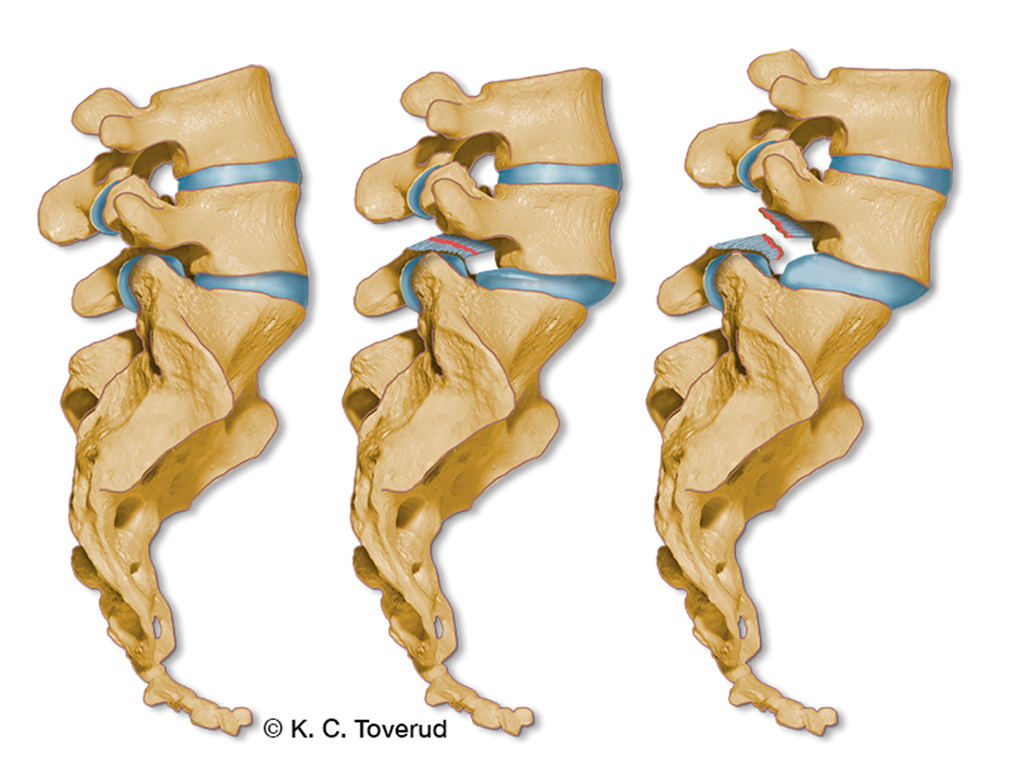
- Spondylolisthesis is a displacement of one vertebra in relation to the next.
- Occurs most frequently between vertebrae L4/L5 and L5/S1.
- Caused by a defect in the pediculus of the vertebral arch
- Degenerative spondylolisthesis is most common and occurs due to age-related changes in multiple structures of the back, as well as changes in the facet joints of the back. This form occurs more often in women, individuals over 50 years old
- Isthmic spondylolisthesis is caused by a defect in the posterior part of the vertebral arch that occurs between the ages of 6 and 16, but is often not recognized until adulthood
- The pain is located in the lower back and can sometimes radiate down the back of the thigh. It is often worst with hyperextension of the back. Sitting often provides relief from the pain
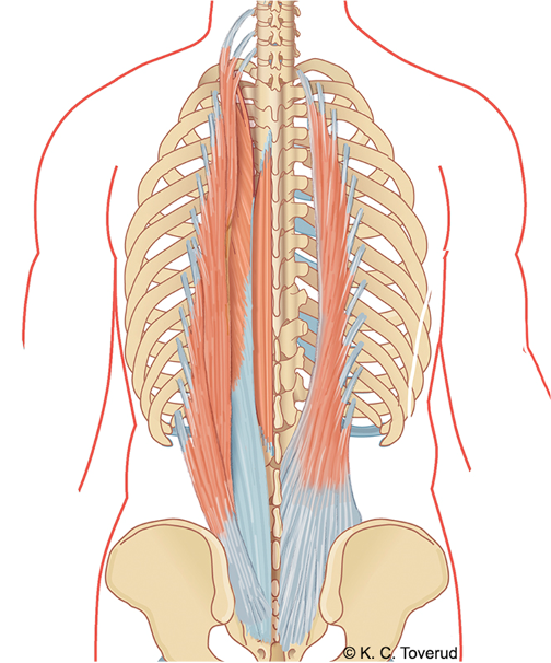
M. erector spinae
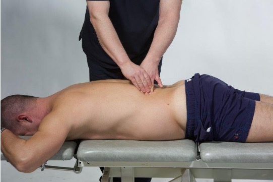
Palpation of m. erector spinae. The patient lies prone. The examiner palpates along m. erector spinae from the lumbar to the thoracic region. Note tensions and pain in the muscles during palpation, and compare with the opposite side.
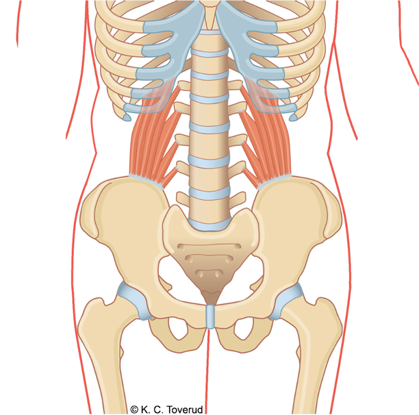
M. quadratus lumborum
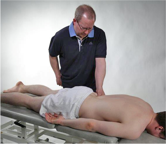
Palpation of m. quadratus lumborum. The patient lies prone. The examiner identifies the highest point of the iliac crest and then moves the hand lateral to m. erector spinae. Then the examiner presses deeper with the thumb of the palpating hand to reach m. quadratus lumborum. Note if palpation triggers pain, and compare with the opposite side.
M. gluteus medius
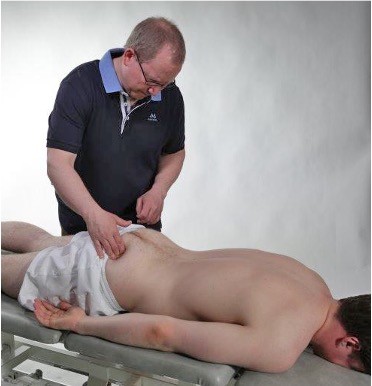
The patient lies prone on the bench. The gluteal muscles are palpated. When palpating the gluteus maximus muscle, you can ask the patient to contract the muscle. Lack of contraction strength if the patient is unable to voluntarily tighten the muscle. Then palpate down towards the greater trochanter and identify the muscle attachment of the gluteus medius and the muscle belly, which is located cranially and somewhat medially to the trochanter. Note if palpation triggers pain, and compare with the opposite side.
M. gluteus medius
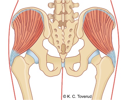
Area of pain in tendinopathy of the gluteus medius

Area of referred pain from tendinopathy in the gluteus medius muscle. Patients with pain over the trochanter area often have signs of tendinopathy in the gluteus medius muscle. Trochanteric bursitis occurs less frequently and almost always in combination with tendinopathy.
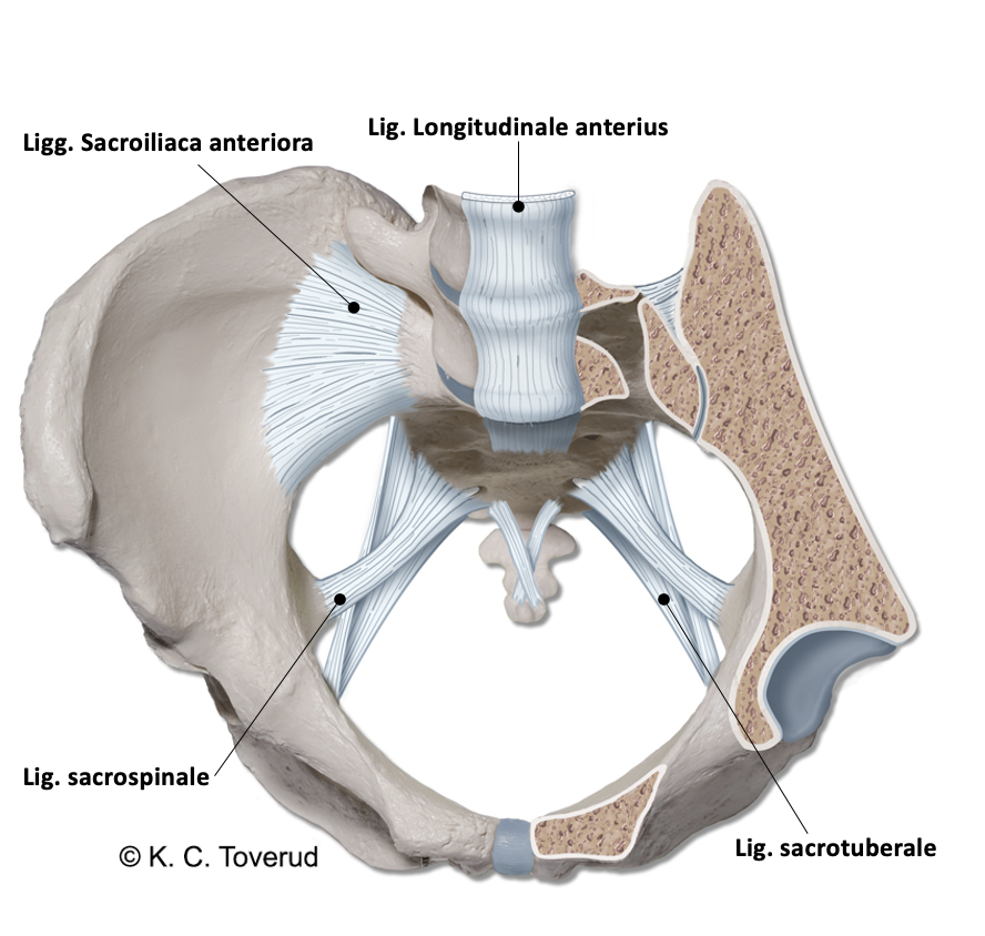
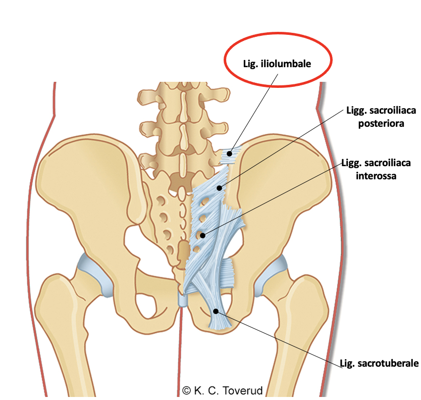
Pain Lig. iliolumbale
- Pain localized from the L4 transverse process down towards the pelvic rim, most commonly due to overuse but also after direct trauma to the area
- Reproduction of pain with specific movements, for example prolonged standing at work or in sports (golf, tennis, volleyball) that involves twisting and hyperflexion/hyperextension movements
- Local tenderness on palpation over the ligament
- Most common in patients with hypermobility in the lumbar spine, marked lumbar lordosis, and reduced strength in core muscles
- Common differential diagnoses are pain from facet joints, spondylosis, myalgia, spondyloarthropathy
Referred pain
Referred pain from structures in the back is common. The pain can originate from costovertebral joints, facet joints, ligaments, intervertebral discs, and the muscles of the back. The distribution of pain can be from the thoracic spine to the chest or from the lumbar spine to the pelvis and thigh.
Assessment of clinical presentation based on functional examination
Pain in the thoracic spine is common. The thoracic spine with ribs is the most rigid part of the spine. In the thoracic spine, weight is transferred from the upper part of the body to the lower parts of the body. Each thoracic vertebra forms between 6 to 13 articular joints. The important joint connections are those between the ribs and the thoracic vertebrae (the costotransverse and the costovertebral joint connections). They form the stability of the rib cage. The extent of joint connections makes it difficult to arrive at specific diagnoses for pain conditions in the joints. Intercostal myalgia and Tietze syndrome can cause chronic pain in the chest, often these are localized and can be mapped by palpation. The most common the location in Tietze syndrome is in the cartilage of the costosternal joint of the 2nd and 3rd ribs. The facet joints in the thoracic spine are oriented so that a large rotational movement can occur here, especially in the middle thoracic vertebrae. Degenerative changes in the facet joints can cause pain during active and passive rotational movement. Pain from the skeleton can also be caused by a thoracic compression fracture after a fall, or pain may arise due to advanced spondyloarthropathy. The functional examination of the thoracic spine primarily aims to rule out that the pain the patient experiences originates from the musculoskeletal system. If negative results are obtained on the functional examination, one must consider note that referred pain may be present. Pain in the thoracic spine can be referred pain from the internal organs, lungs, aorta, and heart. Common conditions that cause thoracic pain are pulmonary embolism, pneumothorax, pleuritis, myocardial infarction, and aortic dissection. Ulcer disease in the esophagus and stomach and gallstones/gallbladder inflammation can give referred pain to the chest. One should also be aware that herpes zoster can cause thoracic pain before skin changes appear.
In the clinical examination of the lower back, one must first determine whether the pain is localized in the lower back or if the lower back pain is associated with radiating pain in the leg. You get an impression of this already during the initial examination of active range of motion in the lower back; pain can be triggered by all movements but most often during extension and flexion. If the pain is localized centrally in the lower back, you need to consider whether the cause is due to degenerative changes in the lower back (vertebral pain) or if the pain is muscular (non-vertebral pain).
By vertebrogenic pain we mean pain caused by changes in bone structures, for example spondylolisthesis, osteoporosis, compression fracture, metastases, or rheumatic disease (for example ankylosing spondylitis). Non-vertebrogenic pain is due to pain in the back muscles, most commonly the erector spinae and deeper muscles, for example the m. quadratus lumborum, which can be palpated to confirm clinical suspicion.
Back pain can also be caused by overstrain of normal ligament structures in the lower back. Usually, pain from ligaments is most often seen in younger people (< 30 years) who complain of diffuse bilateral lower back pain, especially after prolonged standing. There is usually little to be found on clinical examination of these patients.
In some patients with unilateral lower back pain, the cause may be wear in the facet joints, injury to the iliolumbar ligaments, or inflammation in the sacroiliac joint. To get closer to a clinical diagnosis, one must examine the facet joints with the Spring test and the SI joints with sacroiliac joint tests. Injury to the iliolumbar ligaments can be assessed by palpation if the clinical presentation does not suggest that the cause is located in the facet joints or the SI joint.
Some older patients with localized lower back pain may have stenosis, central spinal stenosis, or lateral recess stenosis. In the clinical examination of these patients, there are often few clinical findings; they often have a normal neurological examination. In central spinal stenosis, the patient may report numbness and weakness in the calves when walking (spinal claudication); most often, they experience increased pain during prolonged standing, and on examination, active extension can often provoke symptoms. In lateral recess stenosis, the spinal nerve is compressed in the neuroforamen and can cause unilateral sciatica, few clinical findings on neurological examination, but patients often complain of numbness and "pins and needles." in the calf/foot that can follow the dermatome of the spinal nerve. If there is a disc herniation in the back, most patients will complain of lower back pain with radiating pain in the leg that follows a dermatome. Most patients report pain provocation when sitting, and upon examination, they may often experience worsening of the pain when standing during flexion and extension. Such patients must be examined neurologically with nerve stretch tests (Lasègue and slump test) plus a basic neurological assessment including walking on toes, walking on heels, and squatting. If suspicion of a herniated disc is strengthened through these initial basic examinations, a more detailed neurological examination must be continued. of strength, sensation, and reflexes to clarify the degree of nerve root involvement and attempt to determine which nerve root is most likely compressed.
Special tests
Final competence

- Perform relevant clinical examination in a skilled and gentle manner
- Interpret clinical findings and be able to establish tentative diagnoses
- Suggest referral and treatment
Last updated
31.08.2021