
Inspection
- Assess whether there is joint swelling, and in which joints. Common causes of swelling in the wrist → arthritis or sprain, MCP → synovitis and in PIP/DIP → osteoarthritis (Bouchard's and Heberden's nodes). Assess whether there are contractures (Dupuytren), atrophy (thenar muscles → median nerve compression), ganglion, or deformities due to previous or current fracture.
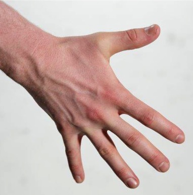
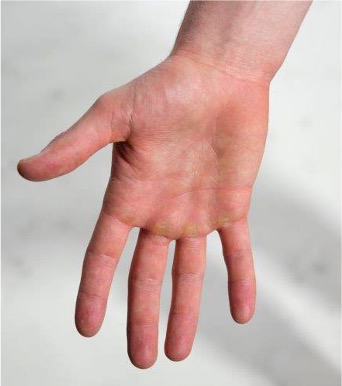
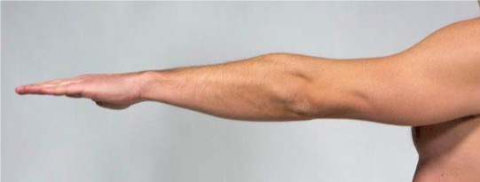
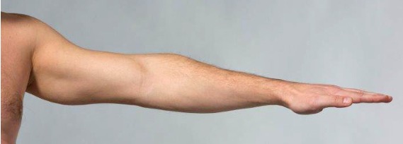
- Consider about whether there are pathological changes in the hand by performing a systematic inspection of the four sides of the hand. The hand is inspected dorsally, volarly, ulnarly, and radially. Look for joint swelling, contracture, muscle atrophy, ganglion, and deformity.
The dorsal side
The volar side
The ulnar side
The radial side
Joint swelling

Assess whether there is swelling in the wrist, MCP, PIP, and DIP joints. Swelling in the wrist is usually caused by arthritis, either as part of an inflammatory disease or following a sprain injury. In the MCP joints, swelling can be due to synovitis, as in rheumatoid arthritis. In the PIP joints, swelling can be caused by Bouchard's nodes, which are bony growths (osteophytes) caused by osteoarthritis. Heberden's nodes are similar changes, but located in the DIP joints.
Dupuytren’s contracture
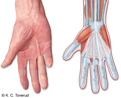
Assess whether there is thickening (fibrosis) in the palmar fascia. Such thickening is particularly found in the palmar fascia over the flexor tendons of the fourth and fifth fingers. This thickening can cause the development of a so-called claw finger, a Dupuytren's contracture.
Contracture in the palmar fascia
Thenar atrophy

Assess whether there is muscle atrophy. Atrophy in the thenar muscles is most commonly seen in carpal tunnel syndrome.
Loss of muscle mass
Ganglion
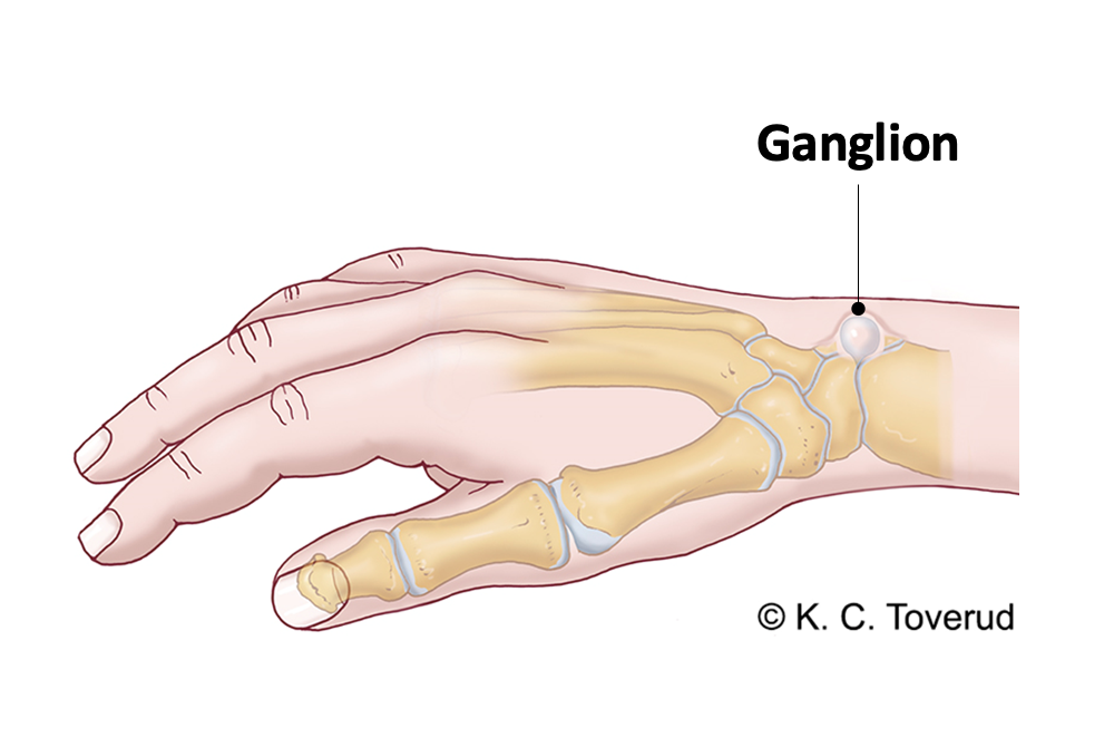
Assess whether there is a ganglion. A ganglion can originate from a joint (wrist or finger joint) or from a tendon sheath. On examination, it is palpated as a cyst-like structure. A ganglion can vary in size over time. It may cause local pain, and in some cases exert pressure on nerves and blood vessels in the hand.
Distal radius fracture
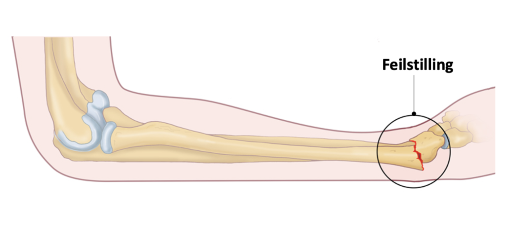
A large proportion of patients with distal radius fractures can be treated conservatively; this applies to those with fractures with no or minimal displacement.
Active movements
- Assess the patient's ability and willingness to move fingers and hand and whether the range of motion is normal, reduced, or asymmetrical. Note whether pain occurs at the start, middle, or end of movement – important to distinguish between tendon, joint, and capsule pathology.
- Consider whether the movement pattern points to flexor tendon injury → lack of active flexion in the PIP/DIP or thumb's IP, extensor tendon injury → lack of active extension in the DIP/MCP, trigger finger → locking during extension from a flexed position, mallet finger → missing active extension in DIP. Also assess whether thumb function (opposition, pinch grip) is weakened (median nerve involvement), and whether pain during active movement corresponds with findings from inspection and palpation.
Wrist flexion
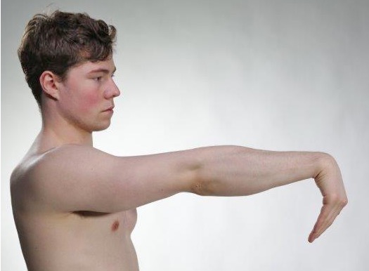
Wrist extension
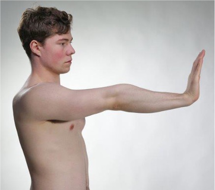
Wrist radial deviation

Wrist ulnar deviation
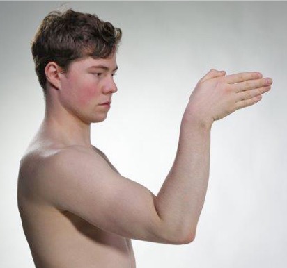
Hand supination

Hand pronation

Finger abduction
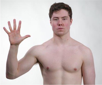
MCP flexion
Extension PIP, DIP
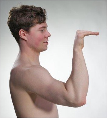
Flexion PIP, DIP
Extension MCP
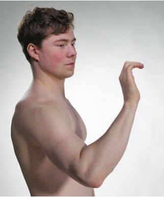
- MCP joint: Articulatio metacarpophalangea
- PIP joint: Articulatio interphalangea proximalis
DIP joint: Articulatio interphalangea distalis - IP joint general: Articulatio interphalangea
Flexor tendon injuries
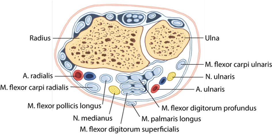
Tendons in the flexor retinaculum of the hand
Testing of the m. flexor digitorum profundus
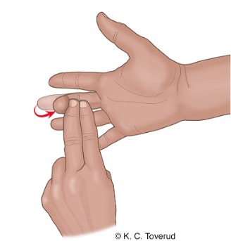
Testing of m. flexor digitorum superficialis
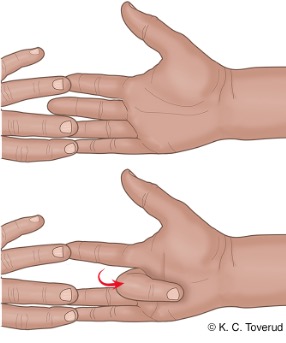
Extensor tendon injuries
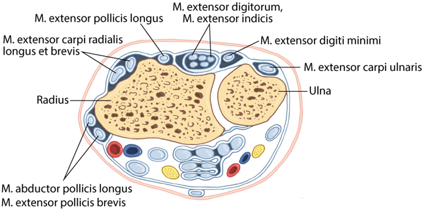
Tendons in the extensor retinaculum of the hand
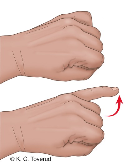
Testing of m. extensor digitorum
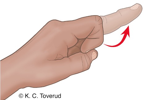
Testing of m. extensor indicis

Extensor tendon injuries
Testing of m. extensor digiti minimi
Trigger finger
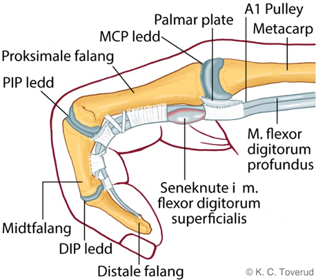
- Trigger finger is caused by the formation of a thickened section on one of the long flexor tendons. When the patient tries to straighten the finger from a flexed position, the thickening gets caught in the opening of the tendon sheath. It is the thickening in the tendon of the m. flexor digitorum superficialis that gets stuck when it tries to pass under the A1 pulley. The thickening is located distal to the pulley. If the thickening were proximal to the pulley, there would be no problem bending the finger.
Mallet finger
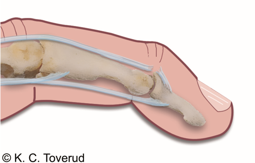
- Mallet finger is caused by an injury or avulsion at the attachment of the m. extensor digitorum on the distal phalanx. The injury results in the inability to perform active extension in the DIP joint.
Thumb flexion
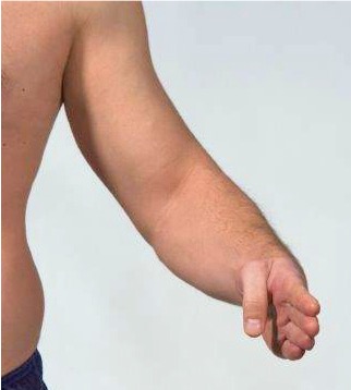
Thumb extension
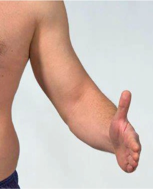
Thumb abduction

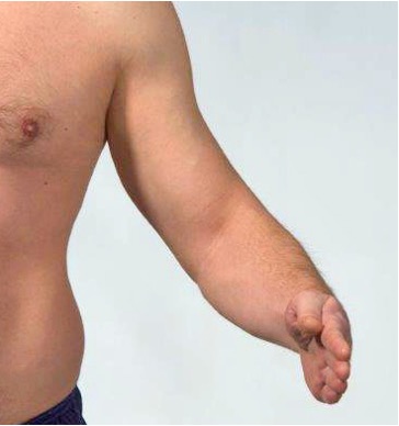
Thumb adduction
Flexor tendon injury in thumb
M. flexor pollicis longus tested by asking the patient to flex the thumb's interphalangeal joint.
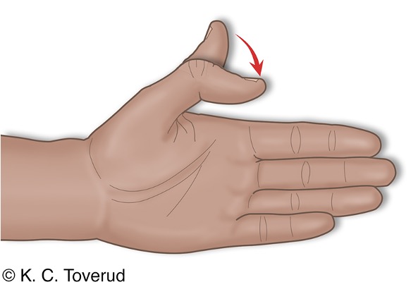
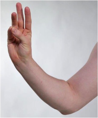
Thumb/little finger opposition grip
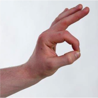
Thumb/index finger pincer grip
Passive movements
- Assess about passive movement is painful, reduced, or has abnormal end-feel. Consider whether the findings point to arthritis → swelling, reduced range, painful end-feel, ligament injury → increased side instability, lack of endpoint in the range of motion, CMC‑1 osteoarthritis → pain with passive extension + adduction of the thumb, crepitation, Skier’s thumb → instability in the ulnar collateral ligament (UCL) of the thumb's MCP, or whether there is de Quervain's tenosynovitis → pain over the radial styloid process with passive ulnar deviation of the wrist.
- Consider About pain during passive movement corresponds with inspection findings (swelling, ganglion, contracture) and findings during active movement. Assess whether the thumb's movements (abduction/adduction) are painful or unstable, which may indicate CMC‑1 osteoarthritis or UCL rupture.
Wrist flexion 80-90°
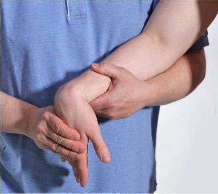
Wrist flexion. Stabilize the forearm with one hand, and flex the wrist passively.
Wrist extension 75-85°
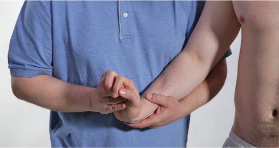
Wrist extension. Stabilize the forearm with one hand, and extend the wrist passively.
Wrist radial deviation 20°
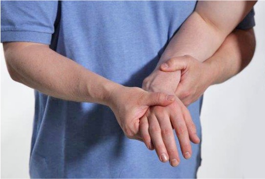
Radial deviation wrist. Stabilize the forearm on the radial side, and move the wrist passively radially.
Ulnar deviation wrist 30-40°

Ulnar deviation wrist. Stabilize the forearm on the ulnar side, and move the wrist passively ulnarly.
Flexion MCP 90°
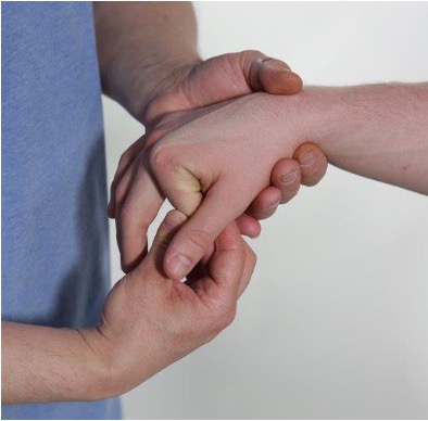
Flexion MCP. Stabilize the wrist with one hand, and move the MCP joint into passive flexion.
Extension MCP 30°

Extension MCP. Stabilize the wrist with one hand, and move the MCP joint into passive extension.
Flexion PIP 120°
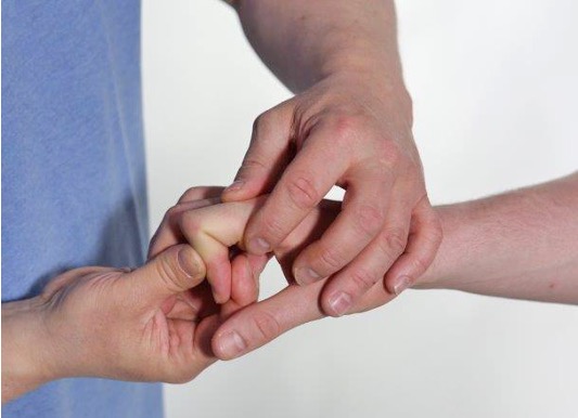
Flexion PIP. Stabilize the MCP joint with one hand, and move the PIP joint into passive flexion.
Extension PIP 10°
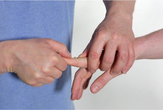
Extension PIP. Stabilize the MCP joint with one hand, and move the PIP joint into passive extension.
Flexion DIP 80°
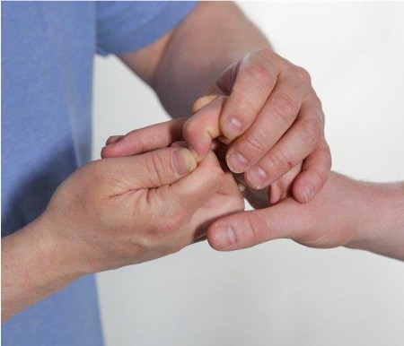
Flexion DIP. Stabilize the PIP joint with one hand, and move the DIP joint into passive flexion.
Extension DIP 10°
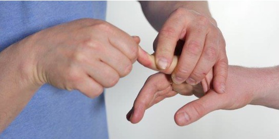
Extension DIP. Stabilize the PIP joint with one hand, and move the DIP joint into passive extension.
Examination of stability in the lateral finger ligaments
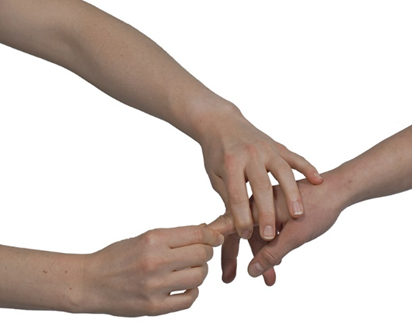
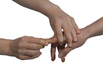
Lateral stability ulnarly in the PIP joint. Stabilize the proximal phalanx radially with one hand, and perform radial deviation in the joint with the other hand. Lateral stability radially in the PIP joint. Stabilize the proximal phalanx ulnarly with one hand, and perform ulnar deviation in the joint with the other hand. Lateral stability in the MCP joint is examined with the joint in full flexion.
Thumb extension 80°
- Carpometacarpal, 70°
- Metacarpophalangeal, 10°
- Interphalangeal, 5°
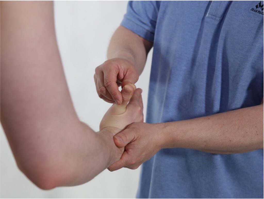
Extension MCP thumb. Keep your wrist in a neutral position, and unevenly stabilize your wrist. Bring the MCP joint of the thumb into full extension.
- Extension thumb 65°
- Carpometacarpal, 15°
- Metacarpophalangeal, 50°
- Interphalangial, 90°
Flexion MCP thumb. Keep your wrist in a neutral position, and unevenly stabilize your wrist. Bring the MCP joint of the thumb into full flexion.
Abduction thumb 60°
Palmar abduction (perpendicular to the palm). Normal range of motion 0–60°
Thumb abduction. Keep the wrist in a neutral position, and stabilize the wrist ulnarly. Abduct the thumb at the MCP joint.
Thumb adduction to 0°
Return of the thumb toward the palm/index finger plane. Normal range of motion is back to 0°
Thumb adduction. Keep the wrist in a neutral position, and stabilize the wrist ulnarly. Adduct the thumb at the MCP joint.
Osteoarthritis CMC1
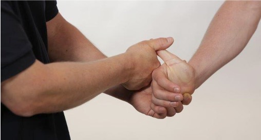
| The CMC 1 joint is prone to the development of osteoarthritis, especially in patients who have hand-intensive work that requires repeated use of strong gripping force in the hand. Clinical examination to determine whether there are signs of osteoarthritis in CMC 1 is done with a pain provocation test where the thumb is moved in a combined motion towards maximum passive extension plus adduction. The movement causes the metacarpal to press against the joint surface, and the examiner notes whether it triggers pain, and if there is crepitus in the joint. On palpation, there will be pain in the joint space, and joint swelling may be present. |
- The CMC 1 joint is prone to the development of osteoarthritis, especially in patients who have hand-intensive work that requires repeated use of strong gripping force in the hand.
Osteoarthritis in CMC1

Ulnar collateral ligament of the thumb

In suspected rupture of the ulnar collateral ligament of the thumb, swelling and tenderness over the ulnar part of the thumb's MCP joint are often found. Clinical examination is performed by fixing the metacarpal with one thumb while the phalanx is radially deviated with a firm grip distal to the MCP joint.
- With clinical signs of instability with > 15° movement, ligament injury is suspected.
Skier’s thumb/Gamekeeper's thumb
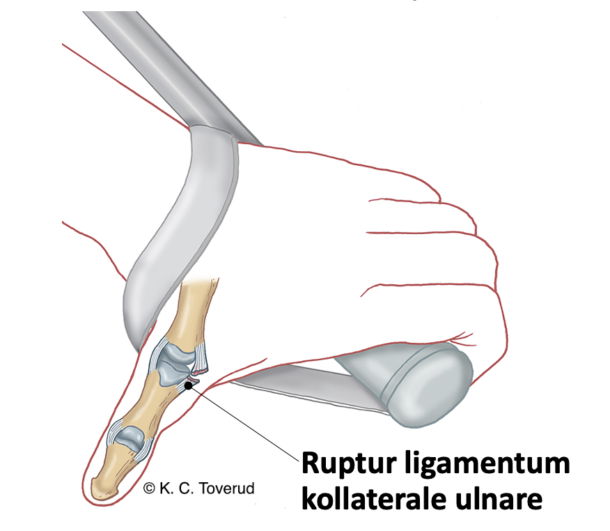
- If instability is found without a clear endpoint, there may be an avulsion fracture at the ligament attachment. An avulsion fracture will be visible on X-ray.
- Partial rupture is treated with immobilization in a cast
- Total rupture is surgically treated and casted
Isometric movements
- Assess Strength in the wrist (extension, flexion), hand grip, pincer grip, opposition grip, finger abduction, finger adduction, thumb (extension, flexion, abduction), little finger (extension, flexion, abduction), and finger (extension, flexion) in MCP, PIP, and DIP.
- Consider Muscle function and differentiate between the different muscle groups.
Wrist radial deviation

Stabilize the forearm with one hand. The wrist is held in a neutral position. Ask the patient to radially deviate the wrist against the examiner's resistance.
Muscles
- m. extensor carpi radialis longus
- m. extensor carpi radialis brevis
Wrist ulnar deviation

Stabilize the forearm with one hand. The wrist is held in a neutral position. Ask the patient to ulnarly deviate the wrist against the examiner's resistance.
Muscles
- m. extensor carpi ulnaris
Interpretation of clinical findings in the wrist

Palpation
- Assess whether there is joint swelling and pain in the wrist, MCP-, PIP- and DIP-joints → possible diagnoses are arthritis, synovitis, osteoarthritis.
- Consider whether palpation for pain and swelling in joints and bone structures reinforces or weakens findings from inspection, active, passive, and isometric movement.
Dorsal wrist

The wrist is palpated dorsally and volarly. Clarify whether palpation triggers pain, and whether there is joint swelling.
Volar wrist

Assessment of clinical presentation based on functional examination
You must first form an impression of whether the range of motion in the wrist is reduced or normal. If the range of motion is reduced, you must determine whether there is a capsular pattern (flexion and extension equally reduced); this could indicate traumatic arthritis in the wrist, early osteoarthritis, or rheumatoid arthritis based on the information you obtain from the medical history. If there is a history of wrist trauma without a previously diagnosed condition, you should consider a missed scaphoid fracture. If, during the examination, you find reduced extension but normal flexion (non-capsular pattern) in a patient with a history of trauma, you should consider whether Subluxation of carpal bones; if there is information about a previous fracture injury, you must consider the development of pseudoarthrosis.
If during the wrist examination you find full mobility and negative isometric tests, you must consider a ganglion in the wrist if the pain occurs when pinching, and an injury to the ulnar or radial collateral ligament if the pain appears during extension.
If, during the wrist examination, you find normal range of motion but pain with extension of the wrist, you must consider that the cause of the patient's complaints may be involvement of one of the extensor tendons (remember the 6 extensor compartments). The most common in general practice is inflammation of the tendon sheath of m. abductor pollicis longus and m. extensor pollicis brevis in compartment 1, so-called de Quervain's tenosynovitis, which you test with the Finkelstein test. Another condition that we can see in general practice is rupture of m. extensor pollicis longus in compartment 3 in connection with undisplaced radius fractures in elderly people. In the other compartments, it is usually tendinopathies or tenosynovitis in patients with acute or chronic overload or in rheumatics.
If during the wrist examination you find normal range of motion, but a positive test with isometric flexion in the wrist, it may be a tendinopathy in m. flexor carpi ulnaris after an acute overload, or a tenosynovitis in mm. flexor digitorum profundus due to overload or as a finding in rheumatics. Be aware that there may simultaneously be clinical findings similar to carpal tunnel syndrome.
Carpal tunnel syndrome causes pain and paresthesia in the median-innervated area of the hand, often in combination with thenar atrophy, tested by Phalen's test and Tinel's sign.
Special tests
Final competence

- Perform relevant clinical examination in a skilled and gentle manner
- Interpret clinical findings and be able to establish tentative diagnoses
- Suggest referral and treatment
Last updated
31.08.2021